Monday, October 10, 2022

How High Is Too High?


Come and listen to a story about a blood test named HDL (High Density Lipoprotein, the “good cholesterol”). It is an epic saga full of lows and highs. It is a story that spans the globe, from Massachusetts to the Italian Alps. What is HDL and how is it associated with heart disease?


The story begins in a town outside of Boston, Massachusetts. In 1977, the famous Framingham Heart Study first identified low levels of HDL in the blood as a risk factor for heart artery disease. In patients with HDL levels less than 40 mg/dl, there is an increased risk for blockage in the heart arteries and cardiac death.  On the other hand, in patients with elevated LDL (the “bad cholesterol”) normal or high levels of HDL protect against heart disease. The prevalence of low HDL in North America is about 7% in men and 2% in women. How does HDL protect against plaque build up in arteries?  There are several mechanisms. HDL transports excess cholesterol from arteries back to the liver, where it is metabolized and excreted. This process is called reverse cholesterol transfer. In effect, HDL “cleans” the arterial wall, sweeping away cholesterol and stopping plaque in its track. In addition, HDL is anti-inflammatory (plaque formation is an inflammatory disease) and has anti-oxidant and anti-blood clotting properties. Are there ways to treat low HDL?  Aerobic exercise, weight loss and smoking cessation all increase HDL levels. Diet plays a role as well. Low fat diets lower both LDL and HDL levels but diets high in monounsaturated fats (including olive oil) reduce LDL without adversely affecting HDL.  Statins will increase HDL levels. They raise HDL between 5% and 15% with an average increase of 9%. Many, many other pharmacologic agents have been tried to see if they can raise HDL levels and improve outcomes. Niacin and fenofibrate both raise HDL (by concomitantly lowering triglycerides). However, despite the positive impact on HDL levels, both drugs failed to reduce heart attacks, strokes and cardiac deaths. Several other medications have been tried, but in clinical trials they all failed to improve cardiac events, some agents even raised mortality. There are several reasons for the failure of theses medications. The biology of HDL appears to be much more complex than that of LDL.  There are several different subclasses of HDL; we don’t know which ones are the keys to the success of HDL. In addition, the function of the HDL molecule is more important than the level. Increasing the amount of HDL doesn’t mean it works better at preventing artery blockage. 


At this point, the story of HDL heads overseas to two small towns in Italy.  In 1980, a genetic mutation in HDL was discovered in families in a town outside of Milan. People with this mutation were smokers, did not follow a heart healthy diet, and had low levels of HDL (10 to 30 mg/dl). Yet they had low levels of blockage in the heart arteries and lived well into their 90’s.  Researchers discovered that their genetic mutation (called apolipoprotein A-1 Milano) was protective against heart disease. Subsequent trials tested whether IV infusions of this apolipoprotein could help patients with established heart disease, but again the trials failed. In another town, this one located in the Italian Alps, there is a cohort of people who have longevity and virtually no heart disease. Like their countrymen, their diet is not heart healthy but they have higher levels of HDL than the average Italian. The genetic variant in this healthy town has not yet been identified.


The guidelines for lipid management recommend a threshold value for HDL of 40 mg/dl; below that level there is an increased risk for heart disease while levels above provide protection. If an HDL value of 40 mg/dl is good, is 100 mg/dl better? Does the risk for heart disease continue to fall as HDL levels rise? How high is too high?  To answer the question researchers pooled multiple studies of HDL (all told more than a million patients were evaluated).  They found a U shaped relationship between HDL levels and death. Levels below 40 mg/dl were associated with increased risk of death. Surprisingly, levels over 80 mg/dl also were associated with increased deaths. They found the optimal range of HDL to be between 40 and 80 mg/dl.


The final chapter to the HDL saga has not yet been written. If you are not genetically gifted and have a low HDL level, do what you can to raise HDL. Stay active, exercise, keep weight down, don’t smoke and take your statin. If you have a very high HDL level, don’t assume that you are protected against heart disease. A healthy lifestyle and a statin may still be necessary if the LDL level is also elevated. Only time and further research will tell whether there is a better approach to HDL and a happy ending to the story. 


Tuesday, September 13, 2022

It Was A Swell Summer


As we say goodbye to the scorching summer of 2022, it is worth noting that heat records have been broken across the globe. Unprecedented heat waves have hit the US, Europe, England and China. According to the National Oceanic and Atmospheric Administration July 2022 was the third hottest July on record, June was the sixth warmest and overall the year 2022 is trending as the third warmest year in history. In the midst of all of this hot humid weather, patients flocked to their doctors with lower leg swelling. What are the causes of leg swelling? Why does it occur with greater frequency in the hot summer months?


To understand why the legs swell, we have to review the blood flow to and the drainage from the lower extremities. The arteries bring blood to the legs from heart. The veins drain the blood back to the heart. There are two venous systems in the legs: the deep veins and the superficial veins. How does blood defy gravity and flow back to the heart? As we walk, the contraction of the muscles in the legs causes the blood to be pumped upward and back to the heart. To facilitate the flow, the veins have valves to prevent blood from leaking back.  Another conduit is the lymphatic system. This consists of thin tubes and nodes that drain lymph back to the heart. Lymph is a fluid that includes excess fluid, proteins, cells, fats and nutrients. About 20 liters of blood flow to the legs every day. The veins drain about 17 liters back to the heart. The lymph system drains the extra 3 liters.  Leg swelling occurs if fluid leaks from the veins or the lymph system because of blockage to the flow or excess pressure within the vessels or the vessels become damaged or dilated. 


Leg swelling can be categorized as acute (recent) or chronic. One leg can be affected (unilateral) or both legs (bilateral).  There are many causes of leg swelling including deep venous thrombosis (a blood clot in the leg, DVT), chronic venous insufficiency, lymphedema, heart failure, kidney failure, liver failure, infection (cellulitis), cancer, thyroid disease, medications, and pregnancy. Medications include calcium channel blockers (especially amlodipine), prednisone, and non-steroidal anti-inflammatory drugs (such as ibuprofen and naproxen).  Leg swelling may be asymptomatic or cause pain, aching, heaviness or fatigue of the leg, skin changes or ulceration of the skin. 


DVT may cause leg swelling in the acute phase as a blood clot could block the flow in the deep venous system. Many patients have swelling years after an acute DVT, likely because the blood clot has caused damage to the venous valves. Treatment of DVT is blood thinners such as warfarin, Eliquis, or Xarelto.


Chronic venous insufficiency is a very common clinical problem. In this condition, the venous valves become incompetent, blood refluxes back into an already congested venous chamber, this increases the pressure in the chamber and fluid leaks from the veins into the surrounding tissue.  Risk factors include older age (peak incidence in women is 40-49 years and men 70-79 years), smoking, pregnancy, hypertension and varicose veins.  Prolonged standing or sitting with the legs in a dependent position also can cause swelling. In this case, the veins are completely filled, the valves float open and fluid leaks out. In addition, warm conditions (such as the hot humid summer weather) tend to aggravate symptoms as the warmth can dilate the veins, causing further venous valvular incompetence.  Conversely, cold conditions relieve symptoms.  The best test to diagnose the cause of leg swelling is a venous ultrasound. The ultrasound can demonstrate a DVT and can show if there is venous reflux/insufficiency.  Chronic venous insufficiency is treated by avoiding prolonged sitting or standing, leg elevation, exercise (walking or calf muscle exercises to help increase the flow) and compression stockings. If swelling persists despite these measures, venous ablation can be performed. Ablation shrinks the refluxing vein, allowing the valves to do their job and decrease or eliminate leaking. Varicose veins are enlarged twisted veins near the surface of the skin. They are usually branches off the superficial or deep vein system. They are quite common (25% of adults have them). Treatment is the same as chronic venous insufficiency but varicose veins can also be surgically stripped or injected with a sclerosing agent to shrivel the vein up. These treatments are more cosmetic than medically necessary. Spider veins are similar to varicose veins, but occur in even smaller surface veins. 


Lymphedema may be primary or secondary to another condition. Primary lymphedema occurs with congenital absence or damage to the lymph system. Secondary lymphedema occurs with blockage of the lymph system due to cancer, prior pelvic surgery, radiation, or trauma. Lymphedema can be differentiated from chronic venous insufficiency by physical exam. In lymphedema there is no pitting of the edema, swelling affects the back of the foot, the toes are involved and there is skin thickening. Lymphedema is usually painless.  Treatment includes compression stockings and pneumatic pump compression. 


So don’t get pumped up over lower leg swelling. See your doctor and get a diagnosis. Then follow the treatment plan to avoid any long-term complications. Keep in mind that cooler weather and relief is coming.


Tuesday, August 9, 2022

What is Optimal Heart Health?


What is optimal heart health? Is it normal heart arteries on cardiac catheterization? Is it the ability to run a marathon? Is it playing a round of golf and walking the course? Is it being able to do moderate exercise without shortness of breath? There are many different and unique perspectives on this question. The American Heart Association’s (AHA) perspective defines optimal heart health using eight variables dubbed “Life’s Essential Eight”.


The AHA initially defined seven variables for optimal heart health in 2010. After 12 years and lots of subsequent research, the AHA updated these seven parameters and added a new one this summer. Here is an update to the original seven with a deep dive into the eighth.

Diet- this was updated to encourage everyone to follow one of the known heart healthy diets; DASH (Dietary Approach to Stop Hypertension) or a Mediterranean Diet.

Physical Activity- no change was made. The optimal level of activity is still 150 minutes per week of moderate physical activity or 75 minutes per week of vigorous exercise.

Nicotine exposure- updated to discourage the use of e-cigarettes (vaping) and to reduce exposure to second hand smoke.

Body Mass Index(BMI) – BMI is body weight divided by height and is a measure of overweight or obesity (A BMI calculator may be found here: https://www.calculator.net/bmi-calculator.html). There was no change to the ideal BMI: between 18 and 25 kg/m2. 

Lipids- the updated metric for blood lipids is non-HDL cholesterol (total cholesterol minus HDL). The ideal non-HDL cholesterol for optimal heart health is < 130 mg/dl.

Blood sugar- optimal levels for non-diabetic patients are a fasting blood sugar < 100 mg/dl or Hemoglobin A1C < 5.7%.

Blood pressure- the optimal blood pressure is less than 120/80 mmHg; 130/80 and above is considered hypertension.


The new variable is sleep duration. According the AHA, the ideal duration of sleep for optimal heart health is between seven and nine hours per night.  The importance of sleep on overall health has been well documented and known for years.  Poor sleep can lead to irritability, anxiety, reduced cognitive performance (including loss of concentration and poor decision making), Alzheimer’s disease and increased risk for obesity, diabetes, heart attack, stroke, hypertension, atrial fibrillation and cancer. Several recent studies outline the risks of not getting a good night’s sleep. A study from the US (6,820 people, mean age 53) looked at various measures of sleep health. Those with poor sleep health had a 54% increased risk for cardiovascular disease. In another study of young healthy people, sleeping only four hours per night added extra weight and specifically added “belly fat”, which has an increased risk for heart disease. The association between sleep and metabolic disease was shown again in a study in women. Sleeping less than 7 hours per night altered glucose (blood sugar) metabolism, leading to diabetes and hypertension. Since sleep is so important, let’s tackle some pressing questions about sleep such as the optimal sleep duration, the best time to go to bed, melatonin and sleep trackers.


What is the optimal amount of sleep? There seems to be a “right” amount of sleep, at least as far as cognitive function is concerned as demonstrated in two recent studies. One study of elderly patients showed dramatic decline in brain function for those who slept less than 4.5 hours or more than 6.5 hours per night. In patients who slept less than 6 hours per night, there was a greater burden of amyloid in the nervous system, a marker for Alzheimer’s disease.  Another study from the United Kingdom (500,00 patients, 38-73 years) showed that seven hours of sleep per night was optimal for cognitive performance. Therefore, seven hours of sleep seems to be the sweet spot for optimal health for middle aged and elderly people (children and adolescents need more sleep). Now that we know the optimal amount of sleep, is there an optimal time for bed?  An interesting study (88,000 patients, average age 61) answers the question. Researchers found that going to sleep before 10 PM or after midnight increased the risk for heart disease. Those who went to sleep between 11 and 11:59 PM had a 12% higher risk for heart disease and those who hit the sack before 10 PM or after 12 AM had a 24% higher risk. The people who went to bed between 10 PM and 10:59 PM had the lowest risk for heart disease in the study; thus defining the optimal time for bed. In this study, timing of sleep was actually more important then duration of sleep. 


If there is difficulty falling asleep and getting the recommended amount of sleep, does melatonin help? Melatonin is a hormone produced by the brain. As darkness falls, the brain senses that it is nighttime and releases melatonin. The elevated levels of melatonin help us fall asleep and stay asleep. As dawn breaks, melatonin levels go down and we wake up. Can over the counter melatonin help people who have trouble falling asleep (insomnia)?  Melatonin has been available for quite some time and is felt to be safe, but no formal studies have been done. Over the counter melatonin is not benign. It has effects on body temperature, blood sugar and blood vessel tone. In addition, since it is not regulated by the FDA, melatonin doses vary widely. The American Academy of Sleep Medicine is currently reviewing the safety and efficacy data of melatonin. Until the review is complete, they recommend that it not be used for insomnia.


Are there devices that can help track sleep? There are a variety of sleep tracking devices that fall into two broad categories: devices put under a mattress or pillow and wearable devices. The devices may help to tell you when you went to sleep and your total sleep time. However, no device can diagnose sleep apnea (stopping breathing). In addition, when compared to a formal sleep study (the gold standard for sleep), no device can accurately measure sleep quality.  In addition, no sleep tracker or sleep app is endorsed for use by national sleep academies or sleep specialists.


So for optimal heart and brain health, go to sleep between 10 PM and 10:59 PM and try to get, on average, seven hours of sleep. Skip the melatonin and the sleep tracker. How many people get enough sleep and score highly on Life’s Essential Eight? It turns out that only one in five US adults (20% of people) have optimal heart health according to the AHA checklist. That certainly is not good; let’s start to improve those numbers. 

Tuesday, July 5, 2022

It's Summer Time!


Summer has arrived. It is time for all of those fun summer activities: golf, tennis, hiking, swimming, kayaking and sitting on the beach. Unfortunately, summer poses some unique risks for the heart patient. How does the heat affect the heart?  Is vacationing good for cardiac health? If so, what type of vacation is beneficial? These questions and more will be answered!

The extremes of temperature, either very hot or very cold, are known to cause additional stress to the heart patient. It is well known that heat waves cause a surge in deaths and hospitalizations for heart disease. Elderly patients are more prone to dehydration which can lead to low blood pressure and increased blood clotting. In addition, very hot temperatures increase the work load of the heart. These effects can lead to a subsequent heart attack or passing out or congestive heart failure. A recent study showed that for each additional extreme heat day (defined as heat index over 90 degrees) there is a 0.13% increase in deaths from heart disease. This translates to an additional 600 to 700 deaths per year in the US due to the heat. With three times as many heat waves per year now compared to the 1960’s, heat related illness must be taken seriously. 

In the absence of a heat wave can a warm summer night be just as risky?  Recent research showed that a rise of just 1.8 degrees Fahrenheit in the usual summer-time temperature caused a 3-5% increase in heart related deaths in men aged 60 to 64, but not in men over 65 or in women. It seems that nighttime temperature had a more potent effect than daytime warmth on death and heart disease. The reason for this is not clear. Socio-economic factors are an issue; those without air conditioning would be more vulnerable to warm temperatures. An intriguing theory involves sleep deprivation. Hot summer nights, especially without air conditioning, could lead to more tossing and turning, interrupting sleep patterns with subsequent increase in blood pressure, heart rate and risk for cardiac events. 

One way to escape the heat is to go on vacation. Can taking a vacation reduce the risk for cardiac events? The Framingham Heart Study showed that men who didn’t take a vacation for many years were 30% more likely to have a heart attack than men who took a yearly vacation. The same study concluded that women who took vacations once every 6 years were 8 times as likely to have heart disease as those who took more frequent time off. The MRFIT trial followed 12,000 men for nine years. It showed that men who took annual vacations were 21% less likely to die and 32% less likely to die from heart disease. Vacationing may have a direct protective effect on heart health for the following reasons. First, vacationing reduces stress and releases one from job demands. Next vacationing may have restorative effects by promoting social contact with family and friends (“reconnecting”). Lastly, certain types of vacations increase physical activity.

So vacations are good for cardiac health. What type of vacation is beneficial; an active vacation or relaxing on the beach? One study from Austria helps answer the question. It showed that an active vacation involving walking, biking or golfing reduced blood pressure and heart rate and helped improve cardiac function. Therefore, an active vacation may improve cardiac health more than lying in the sun. It seems that physical activity during leisure time (including vacations) is beneficial. How does physical activity during leisure time compare to physical activity at work?  The Copenhagen General Population Study followed 104,000 people over 10 years and had an interesting answer to the question. The study found that physical activity during leisure time reduced the risk for cardiac events by 15% and the risk for death by 40% but physical activity at work increasedthe risk for cardiac events and death. They called this the “physical activity paradox”. Why is this? They theorized that leisure time activity was more dynamic, more likely to improve cardiorespiratory fitness and allows sufficient recovery time. Physical activity at work, on the other hand, is more static, monotonous, often occurring with awkward positioning and did not allow for meaningful recovery time. 

So as we head into summer, make those vacation plans. Then dust off the golf clubs or hiking boots. Lastly, crank up the air conditioning at night and get a good night’s sleep.


Monday, June 6, 2022

The Calcified Aortic Valve


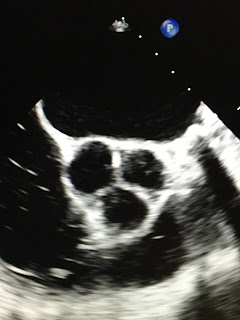
The aortic valve is a three-leaflet valve situated between the left ventricle (the main pumping chamber of the heart) and the aorta (the main artery leading from the heart). When the left ventricle contracts to send blood to the body, the aortic valve opens to allow the blood to flow out. After the blood is ejected, the valve closes to prevent blood from leaking back in to the heart. The aortic valve opens and closes, under a tremendous amount of pressure, with each heartbeat, for our entire lives. Because of this, the aortic valve is subject to “wear and tear” over time. As the valve wears down, the leaflets become thickened and calcium is deposited to repair microtears in the valve.  As the valve becomes more thickened and calcified, it doesn’t open fully, restricting the blood flow from the heart. This pathologic condition is called calcific aortic stenosis. When there is significant restriction of the blood flow, it is called severe aortic stenosis. Severe aortic stenosis may cause chest pain, passing out, congestive heart failure or sudden cardiac death. Unfortunately, once severe symptomatic aortic stenosis occurs, there is no medication to treat it. The only way to restore blood flow is to replace the valve. This is done by surgically removing and replacing the aortic valve or, more recently, the valve can be replaced without surgery by inserting a new valve through the artery in the groin (TAVR). Currently there are no medications that can prevent calcific aortic stenosis or slow its progression once it occurs. Or is there?


Calcific aortic stenosis is very common occurring in 2% of people over the age of 65 and 4% over 85 years old. It is associated with older age and shares other risk factors for atherosclerosis (blockage in the heart arteries). It has been known for some time that plasma lipids (cholesterol) are involved in the process of thickening and calcification of the aortic valve. It seems reasonable to presume that if elevated cholesterol is a risk factor that lowering cholesterol with a statin would halt the process. Many studies with statins have been performed. Unfortunately, statins do not halt the progression of severe calcific aortic stenosis. What if a statin is started before the disease is far advanced? Again, statins did not slow the progression in patients with mild aortic stenosis.  So, lowering cholesterol doesn’t work. Are there other targets? One possibility is PCSK9. PCKS9 is a protein involved in the production of LDL cholesterol. In patients with heart artery disease PCSK9 inhibitors (such as Praluent or Repatha) dramatically lower LDL cholesterol and reduce the risk for heart attack and cardiac death.  Recently it has been shown that patients with calcific aortic stenosis have higher levels of PCSK9. It is felt that PCSK9 plays a role in mediating aortic stenosis through inflammation and the stress responses of the cell. Do PCSK9 inhibitors slow the progression of calcific aortic stenosis? We don’t know, but future trials may give an answer. Another possible target is lipoprotein a. Lipoprotein a is an LDL like particle that is also associated with elevated risk for heart artery disease. Elevated levels of lipoprotein a have been shown to correlate with progression of calcific aortic stenosis as well. Patients with the highest level of lipoprotein a were twice as likely to need aortic valve replacement as patients with low levels of the protein. Statins do not lower levels of lipoprotein a, but PCSK9 inhibitors do. Whether this translates to a beneficial effect for calcific aortic stenosis is not known, but it seems a trial with these agents is badly needed. 


Another potential target is cardiac amyloid. What is amyloidosis? The pathogenesis of amyloid involves a protein called TTR which is produced by the liver and aids in transporting thyroid hormone. In some people (older patients, patients with genetic predisposition), TTR clumps together to form amyloid fibrils. These amyloid fibrils are deposited in many tissues in the body, but especially the brain, the nerves and the heart.  Amyloid deposition in the heart can cause a variety of problems including congestive heart failure as well as calcific aortic stenosis. In patients older than 75 years old with aortic stenosis, 14% have cardiac amyloid.  Cardiac amyloid is treated with a medication called Tafamidis, which stabilizes TTR and prevents the formation of amyloid fibrils. Tafamidis slows the progression of cardiac amyloid in patients with congestive heart failure, but it is not known whether it does the same for calcific aortic stenosis. This too may be a promising area and ripe for future research. 


Another potential target is calcium. Since calcium is deposited in the aortic valve do calcium supplements increase the risk? A study of 2600 patients with mild to moderate aortic stenosis found the following. Aortic valve replacement was ultimately needed in 50% of patients taking calcium supplements versus only 11% in patients not taking supplements. It was concluded that long-term calcium supplementation should be avoided in calcific aortic stenosis. 


If you have calcific aortic stenosis, don’t despair. A newer and better treatment (TAVR) has already dramatically changed the life for many a patient with this disease.  Although we currently cannot stop calcific aortic stenosis or alter its progression, newer targets and newer treatments may be on the horizon offering similar dramatic medical options to improve the lives of patients. In the meantime, hold those calcium supplements. 


Tuesday, May 10, 2022

April Showers Bring May Green Prescriptions


As the rain in April turns into the beautiful trees, shrubs, grass and flowers of May, we can see that green is good!  As we sit gazing out of our window at the marvelous greenery, we wonder, “Is it better to exercise indoors in a gym or outdoors in nature? How else is green good for health?” The US Environmental Protection Agency has estimated that Americans spend 90% of their time indoors. Is this healthy? Should we heed our mothers’ advice and “Go outside and get some fresh air”?


It has been felt for a long time that getting back to nature, spending time in natural environments, had positive mental and physical effects.  Being outside supposedly lowered blood pressure, decreased obesity, helped with diabetes, reduced anxiety and depression, lessened stress, and promoted physical activity while combating sedentary leisure time. Because of these effects, doctors have given out nature prescriptions, essentially encouraging patients to get outside. There is even a Japanese practice called “forest bathing” (shinrin-yoku) whereby health benefits are obtained just by sitting in nature. Is there any data to support these health claims? One study of forest bathing showed that the practice increased natural killer cells and other cancer fighting proteins. Researchers theorize that the trees emit compounds that help plants fight disease, but apparently also help humans fight cancer as well. Another study showed that exercising in natural environments reduced stress, decreased anger, lessened depression and improved energy compared to exercising indoors. A different study compared a three-hour mountain hike with walking on treadmill indoors and with sedentary activities. Mountain hiking improved a variety of psychological parameters compared to the treadmill and sitting indoors.  A 2019 study looked at nearly 20,000 people in England and found that spending two hours per week in green spaces (parks or other natural environments) improved people’s health and psychological well-being.  The two hours could be all at once or spread over the week.  The two hours also held for those who were ill or disabled, suggesting the time in nature was the key rather the effect of healthier people outside exercising more often.  It seems that exercising outside, in a natural green space, is better than indoors. Even if the data isn’t entirely true, there is no downside risk to being outside except for encountering a few raindrops now and then.


How about living near green spaces? Is there a health benefit? One study looked at the green space of Toronto.  They found that people who live in areas with more trees had less hypertension, diabetes, heart attacks and stroke. They found that having 11 or more trees in a city block reduced the risk for cardiac disease.  In addition, researchers looked at NASA images of the United States and categorized each county’s greenness (a measure of trees, shrubs and grass). For every 0.1 unit increase in greenness, deaths from heart disease fell by 13 deaths per 100,00 people. This was confirmed in a study from Spain.  People who live near green spaces had a 16% reduction in their risk for stroke. The mechanism of benefit of green spaces may be the lower amount of air pollution and better air quality in general in these areas. Green is good!


Another type of green is good for overall health. Green leafy vegetables (for example, arugula, green beans, kale, spinach, asparagus, broccoli, zucchini) have significant health benefits.  Three servings a day reduced the risk of diabetes by 13% and lowered the risk for heart disease by 24%. Now there is new information about a different green. Add avocado to the list of healthy greens. People who ate two avocados per week had a 22% lower risk for developing cardiovascular events.


How can we put all this greenness to work for us? With a Green Prescription. The prescription itself is more than just a ticket to get medication at the pharmacy. It is a contract between the doctor and the patient. With that in mind, here is your contract with your doctor and with nature, the Green Prescription: 

1) Spend 120 minutes per week outdoors in nature

2) Eat 3-4 servings of green leafy vegetables per day

3) Have 2 avocados per week

Munching on an avocado and some celery sticks while hiking through the forest is optional.


Tuesday, April 5, 2022

The Intersection of Breast Cancer and Heart Disease

(Google Images)

A 69-year-old woman presents to her cardiologist with shortness of breath.  She was diagnosed with congestive heart failure (CHF).  An echocardiogram showed an ejection fraction (a measure of the pumping function of the heart) of 25%, representing a severe weakness of the heart muscle. Her past medical history is remarkable for left sided breast cancer diagnosed in 2000. She was treated with lumpectomy and chemotherapy.  She was felt to be cancer free in 2008. What caused her CHF and her weak heart?


Breast cancer is the most common form of cancer in woman in the United States. Approximately 1 in 8 women will develop breast cancer.  Breast cancer does occur in men as well, although it is rare, accounting for only a few cases per year.  Breast cancer is treated with surgery, radiation therapy and chemotherapy.  Fortunately, cancer treatments are curing more and more breast cancer. Unfortunately, those same treatments can cause heart disease, often years later.  Radiation, especially to the left side, can affect all of the layers of the heart. It can affect the outer layer of the heart causing inflammation and chest pain (pericarditis). Radiation can damage the heart muscle and cause it to weaken. Lastly, radiation can cause blockage in the heart arteries. Studies have shown that patients who had radiation for left-sided breast cancer are twice as likely to have heart artery disease as patients with right-sided cancer.  Radiation can accelerate blockage starting about 5 years after therapy and the effect may persist for up to 30 years. Chemotherapy agents are very good at inducing cures, but two agents in particular can cause significant heart damage. Adriamycin acts as a toxin to the heart muscle. It causes weakening and a reduction in the ejection fraction, leading to CHF, often 10 to 20 years after the chemotherapy is given. Unfortunately, Adriamycin’s effect is irreversible. Another agent, Herceptin (Trastuzumab) also causes a weak heart muscle and CHF. However, stopping the agent does allow the heart to recover function, so vigilance is needed during its administration. 


Breast cancer and heart disease share a number of risk factors including: older age, poor diet, alcohol use, obesity, sedentary lifestyle and smoking. In a two for one deal, exercising, keeping weight down, not smoking and limiting alcohol not only reduces the risk for heart disease, but also lowers the chance of getting breast cancer. This advice is valid for both women and men (remember, men can get breast cancer as well). Two strategies in particular are worth noting for their reduction in both heart disease and breast cancer: exercise and taking a statin for cholesterol.  Exercise capacity has shown to be a prognostic factor in breast cancer.  Patients with higher levels of physical activity have a lower risk of developing breast cancer and dying from the disease.  Patients with better cardiorespiratory reserve, developed from years of exercising, can tolerate the toxic effect of chemotherapy. These patients suffer less cardiac side effects during treatment. In addition, these patients are more likely to stay active during chemotherapy, thus lessening their risk further. Statins may be protective as well. Women who were taking a statin during chemotherapy had a lower risk for CHF. This may be because, beyond lowering cholesterol, statins decrease inflammation and stress in heart cells. 


Screening for breast cancer may be a useful tool for screening for heart disease too. The mammogram (an X-ray of the breast) is the standard test for screening for and detecting breast cancer.  If a suspicious area is seen on mammography, then a biopsy is done to confirm or rule out cancer.  Breast artery calcification may also be seen on mammography. Breast artery calcification, like heart artery calcification, signifies the presence of plaque or blockage in the arterial wall.  Heart artery calcification on CT scan is associated with a higher risk for heart attack and heart death and a reason to intensify preventive treatments.  Does breast artery calcification have the same prognostic significance? In a study of 5000 women (all over age 60) who did not have breast cancer or heart disease, 26% were found to have breast artery calcification. Those with calcification were twice as likely to have heart disease or stroke. In another two for one deal, it looks like mammography can screen for bother breast cancer and heart disease. 


The index case and the three intersections between breast cancer and heart disease teach us the following lessons. Patients who have had breast cancer and who had radiation or certain types of chemotherapy must remain vigilant about heart symptoms and see their doctor if symptoms do occur.  With common risk factors, following a healthy lifestyle reaps rewards in numerous ways. Lastly, artery calcification anywhere in the body must be taken seriously and preventive medications must be initiated.  Following these measures can help avoid two deadly diseases: breast cancer and cardiovascular disease.