Thursday, September 19, 2019

A Bayesian Approach to the Diagnosis of Hypertrophic Cardiomyopathy During Athlete Screening

Screening athletes for causes of sudden cardiac death (SCD) is difficult. The conditions that cause SCD are rare and sometimes difficult to diagnose, especially in athletes under 35 years old. Many hundreds of athletes must be screened to find even a single case of a potentially lethal heart condition. In many series of SCD in athletes, hypertrophic cardiomyopathy (HCM) is a leading cause (1). There are characteristics of HCM that can be detected by the history (syncope, family history), physical exam (a systolic murmur louder on Valsalva maneuver) and by electrocardiogram (EKG).  In addition, it can be detected easily on echocardiography (echo), even a quick screening echo.  Therefore, since HCM can be deadly and since it can be detected using information available at a routine screening, the diagnosis must be made. Would an additional tool, such as Bayesian analysis, be helpful in diagnosing HCM during an athlete screening session?

Bayes Theorem is useful when trying to make a diagnosis under uncertainty.  It closely follows clinical medicine; as information is received, the probability of the diagnosis is revised either upward or downward. Bayes Theorem requires three inputs: a pretest probability (or prevalence of disease), the sensitivity of a finding or a test and the specificity of the finding or test.  It works as follows. A patient comes to a doctor’s office and a disease is suspected. The history is taken and the patient reports a symptom. The doctor knows the prevalence of the disease in the population and looks up the sensitivity and specificity of the finding in picking out the disease entity.  The numbers are put in Bayes formula and a posttest probability is calculated. In other words, the patient is suspected of having a disease, reports a symptom consistent with the disease and the doctor’s suspicion of the disease then increases. If the patient has a physical finding consistent with a disease, then the posttest probability just calculated now becomes the pretest probability and the sensitivity and specificity of the physical finding are used to find a new posttest probability.  If the patient then has an abnormal EKG, then previous posttest probability and the sensitivity and specificity of the EKG abnormality are used to revise the estimate of disease probability, and so on.  As new information is obtained, the previous probability of disease is used to come up with a new estimate- exactly as is done in an office setting- as new information comes to light, the probability of disease goes either up or down. This approach was used by Diamond and Forrester (2) to calculate the probability of coronary artery disease and has been used many times since then. This same method can help diagnose HCM during screening of athletes.

The prevalence of HCM is well known (3). The generally accepted prevalence is that 1 person out of 500 people (0.2%) in the population will have HCM.  This has recently been revised and the new estimate is 1 person out of 200 people (0.5%) may have HCM.  For the purposes of the screening tool, both numbers are used to provide a range of probabilities. 

A detailed history and physical are the cornerstones of the cardiovascular screening of athletes. Every athlete fills out a standard American Heart Association questionnaire and a physical examination is performed.  For the purpose of diagnosing HCM, two items on the questionnaire are of interest.  A prior history of unexplained syncope may be associated with HCM.  This has been studied and it has been determined that the sensitivity for unexplained syncope in diagnosing HCM is 35% and the specificity is 85% (4). Since HCM is a genetic disease and runs in families, the family history is very important.  A family history of unexplained SCD has a sensitivity of 42% and a specificity of 79% in diagnosing HCM (4).  A family history of HCM carries a sensitivity of 44% and a specificity of 99% (5). Findings on physical examination can also determine the presence of HCM.  The classic murmur of HCM is a harsh systolic murmur that gets louder with Valsalva maneuver. The sensitivity of a systolic murmur, louder with Valsalva, is 65% while the specificity is 96% (4). The history and physical examination may not be able to definitively diagnose HCM (the sensitivities are quite low), but if these factors are present, the probability of HCM increases and additional testing is warranted.

The next test during an athletic screening is the EKG.  While controversial and not performed routinely in all parts of the world, the EKG should be done if one suspects HCM.  Many patients with HCM have abnormal and bizarre EKGs.  An EKG is abnormal if it meets the findings of the 2013 Seattle criteria and the updated 2017 International criteria.  The sensitivity of an abnormal EKG in diagnosing HCM using the Seattle/International criteria is 93% with a specificity of 96% (6).  

Lastly, an echo is often done during screening of athletes.  Usually an echo is performed if there is a reasonable probability that a condition which may cause SCD is present.  An echo is often used to rule in or rule out a diagnosis of HCM.  While the differentiation between HCM and an athlete’s heart on echo can be difficult, at screening one needs to determine if the heart is hypertrophied or not and whether additional testing is necessary.  There are many criteria used to diagnose HCM on echo, but three criteria are the generally accepted starting points in making the diagnosis: interventricular septal wall to posterior wall ratio greater than or equal to 1.3, systolic anterior motion (SAM) of the mitral valve and maximal interventricular septal thickness >1.5 cm (7).  These three parameters are easily obtained on echo during a screening session for athletes; they don’t require additional expertise by the echo tech or echo reader. An interventricular septal wall to posterior wall ratio greater than or equal to 1.3 has a sensitivity of 76% and a specificity of 93% (7). Systolic anterior motion of the mitral valve has a sensitivity of 82% and a specificity of 99% (7). Maximal interventricular septal thickness >1.5 cm has a sensitivity of 87% and a specificity of 97% (8). 

Table of Sensitivities and Specificities in Diagnosing HCM
Sensitivity
Specificity
Unexplained syncope
0.35
0.82
Family History of unexplained SCD
0.42
0.79
Family History of HCM
0.44
0.99
Systolic murmur increased w/Valsalva
0.65
0.96
Abnormal EKG - Seattle/International Criteria
0.93
0.96
Septal/Posterior wall ratio => 1.3
0.76
0.93
SAM
0.82
0.99
Interventricular septum > 1.5 cm
0.87
0.97
                                                                                                            

How does the Bayes calculator work? Currently, it is a spreadsheet and the relevant factors (ex, family history HCM) are set to a default of 0.  If a factor is positive, then the 0 is replaced by a 1 and a new posttest probability is displayed.

Take for example an athlete whose only positive finding is a prior history of unexplained syncope, the physical is normal and the EKG is normal. In this case, the baseline probability of HCM goes from 0.2% - 0.5% to 0.4% - 1%. This is still quite a low probability and if one is wondering whether to do an echo, it may acceptable to skip additional testing. 
1/500
1/200
Prevalence of HCM
0.002
0.005
Unexplained syncope
1
1
Family History of unexplained SCD
0
0
Family History of HCM
0
0
Systolic murmur increased w/Valsalva
0
0
Abnormal EKG - Seattle/International Criteria
0
0


Post Test Probability
0.004
0.010


Now if the same athlete has a history of unexplained syncope and an abnormal EKG by Seattle/International criteria, then the probability of HCM goes to 8%- 18%. This is now in an intermediate range and doing an echo would be prudent.


1/500
1/200
Prevalence of HCM
0.002
0.005
Unexplained syncope
1
1
Family History of unexplained SCD
0
0
Family History of HCM
0
0
Systolic murmur increased w/Valsalva
0
0
Abnormal EKG - Seattle/International Criteria
1
1


Post Test Probability
0.083
0.185

If an echo is done and the interventricular septum is greater than 1.5 cm, the posttest probability goes to 72-87%, all but cinching a diagnosis of HCM.

1/500
1/200
Prevalence of HCM
0.002
0.005
Unexplained syncope
1
1
Family History of unexplained SCD
0
0
Family History of HCM
0
0
Systolic murmur increased w/Valsalva
0
0
Abnormal EKG - Seattle/International Criteria
1
1


Post Test Probability
0.083
0.185
Echo
Septal/Posterior wall ratio => 1.3
0
0
SAM
0
0
Interventricular septum > 1.5 cm
1
1
Post Test Probability (+ echo)
0.724
0.868
Post Test Probability ( - echo)
0.016
0.040


The calculator may be helpful in another way. For example, take an athlete who has a family history of HCM and an abnormal EKG. The posttest probability is quite high (67-84%). If, however, the athlete has a negative echo, the posttest probability of a negative echo drops to 27 to 48%. While the probability at this time is lower, this athlete should not be ignored, the probability of HCM is still close to 50%. This scenario represents an athlete who should be followed over time and have repeated echoes.  Even though he doesn’t have HCM at present, he is at risk and may develop it in the future. Young athletes with abnormal EKGs and normal echoes may represent an early phase of the disease and it may be evident years later (9). The calculator may help identify these athletes.

Other causes of SCD in athletes are even less prevalent than HCM. In addition, they do not have the same clues on history and physical examination. The EKG can have subtle abnormalities or it can be normal. The echo can be diagnostic, but oftentimes additional testing (stress testing or cardiac MRI for example) is necessary to diagnose one of these conditions. These tests are not part of a screening evaluation for athletes. Take arrhythmogenic right ventricular dysplasia (ARVD) as an example. The prevalence is 1 in 5000 (ten times higher than HCM) (10). Sensitivities and specificities are known for common EKG abnormalities in ARVD (11).  The pretest probability of ARVD is very low due to the low prevalence, about 0.02%. Even if the EKG is abnormal, the posttest probability is only 1.3%, still quite low. The Bayesian approach may not be helpful.

ARVD
1/5000
Prevalence
0.000200
Positive Family History- ARVD in first degree relative
0
Inverted T waves in V1, V2 and V3 or beyond in patient > 14 yrs old, w/o RBBB
1
Epsilon wave in V1, V2, V3, w/o RBBB
1


Post Test Probability
0.013598

Using the Bayes calculator may be helpful in diagnosing HCM during a routine athlete screening and in identifying athletes who need to be followed closely over time.  The tool uses commonly available variables during a typical athlete screening session and takes only a few minutes to use. However, it has not been prospectively validated. 

Steven Georgeson, MD FACC FACP
Medicor Cardiology
Atlantic Medical Group
Bridgewater NJ USA
References:
1.    Circ 1996;94:850-856
2.    NEJM 1979;300:1350-1358
3.    NEJM 2018;379:655-668
4.    Heart 2004;90:570-575
5.    Am J Card 2014;114:1383-1389
6.    Br J Sports Med 2018;52:667-673
7.    JACC Imaging 2008;1:787-800
8.    JACC 1993;22:498-505
9.    NEJM 2008;358:152-161
10.NEJM 2107;376:61-72
11.Circ 2009;120:477-487


Monday, September 9, 2019

The Magnificent Seven


Today we pay homage to the number seven. The number is seven is special, cropping up in diverse aspects of life and across the years of history. For example, one can sail the seven seas. There were seven wonders in the ancient world. “The Magnificent Seven” was a movie from 1960, one of the best westerns of all time. “The Magnificent Seven” is also the name of a song by the Clash, chronicling the seven hours of the workday in London in the 1980’s. Seven-card stud is a form of poker. Mickey Mantle wore number seven! How is seven significant in the cardiology realm?

In 2010, the American Heart Association (AHA) defined and set national goals for cardiovascular health. The goal, “By 2020 to improve the cardiovascular health of all Americans by 20% while reducing deaths from cardiovascular disease and stroke by 20%”.  The AHA defined seven health factors or behaviors that were associated with cardiovascular health. The AHA called these factors Life’s Simple 7 and they are: physical activity, blood cholesterol, healthy diet, blood pressure, healthy weight, blood glucose(sugar) and smoking.  Each factor was broken into three categories: parameters associated with ideal cardiovascular health, parameters associated with intermediate cardiovascular health and those associated with poor cardiovascular health.  In addition, points are awarded for falling into each category: 0 points for poor health, 1 point for intermediate and 2 points for ideal health.  Here are the categories:

Health Factor or Behavior
Poor Cardiovascular Health
(Warning)


(0 points)
Intermediate Cardiovascular
Health
(Needs Improvement)

(1 point)
Ideal Cardiovascular Health
(Excellent)


(2 points)
1. Physical Activity
Little to none
1-149 min/week moderate exercise or < 74 min/week vigorous exercise
150 or more min/week moderate exercise or 75 or more min/week vigorous exercise
2. Cholesterol
=> 240 mg/dl
200-239 mg/dl
or treated to goal
< 200 mg/dl
3. Healthy Diet
Portions per day:
   5 cups fruit/vege
   4 oz whole wheat
   < 1500 mg of            sodium

Portions per week:
   2-3 servings fish
   < 450 calories       from sugared drinks
0-1 components
2-3 components
4-5 components
4. Blood Pressure
Systolic => 140
Diastolic => 90
Systolic 120-139
Diastolic 80-89
Systolic < 120
Diastolic < 80
5. Healthy Weight
Body mass index (BMI)
BMI => 30
BMI 25-29.9
BMI < 25
6. Blood Glucose
Blood sugar while fasting
=> 126 md/dl
100-125 mg/dl
< 100 mg/dl
7. Smoking
Current smoker
Quit < 12 months ago
Never smoked or quit > 12 months ago


It is now nine years into the AHA campaign. Has there been an impact from Life’s Simple 7?  The overall rates of heart disease and stroke have been declining for a number of years. However, the death rates for heart disease and stroke, which had been declining as well, have leveled off and may even be increasing. Some of this may be explained in the context of Life’s Simple 7. The percentage of adults who meet ideal status for smoking, blood pressure and cholesterol have been increasing. Unfortunately these gains are offset as the percentage of adults whose BMI and glucose in the ideal range have been decreasing (leading to the obesity and diabetes epidemic).  In fact the prevalence of obesity increased from 22% in the 1990’s to 35% in 2012, while diabetes tripled (2.5% in 1990 to 7.2% in 2013). However, following Life’s Simple 7 can improve longevity. One study followed 7600 adults for about 6 years. The study found that participants who met 5 or more of the ideal metrics had an 88% reduction in heart deaths compared to those who met none of the ideal metrics. An analysis from 2014 showed that heart disease improved by only 6% since 2010, far short of the goal of 20% reduction by 2020. Yet following Life’s Simple 7 can help keep the heart strong. Many studies have looked at congestive heart failure and Life’s Simple 7.  A healthy lifestyle score was developed summing the points in each category (the range is from 0 to 14).  An inadequate score was 0 to 8, an intermediate score was 9 or 10 and an ideal score was 11 to 14.  Participants with an intermediate score had a 47% lower chance of congestive heart failure compared to those with an inadequate score. Individuals with an ideal score had a 55% lower chance of heart failure. In addition, those with intermediate and ideal scores had better preservation of the heart’s structure and function (lower risk for a thickened heart and weakening of the heart muscle).

It certainly seems that adhering to the lifestyle promoted by Life’s Simple 7 will go a long way towards reducing heart disease and improving longevity.  So, don’t gamble with your life. Don’t bet on the cards falling your way. Instead, follow Life’s Simple 7 (or the Magnificent Seven) for years of a heart healthy life. 

Monday, August 5, 2019

An Apple or a Pear?


At the food court in the mall there are three people in line waiting to buy pizza. The first person in line is a man who is 6 feet tall and weighs 250 pounds. He is very muscular and has no discernable body fat. The second is a woman who is 5 feet 2 inches tall and weighs 131 pounds, but has a lot of belly fat.  The third person is a woman, 5 feet 6 inches tall and 149 pounds, with heavy legs.  What is the risk for heart disease for these three people?

Obesity is a well-known risk factor for heart disease. There are a variety of ways to measure obesity. The body mass index  (BMI, derived from a formula using weight in pounds and height in inches) is the standard measure used to define whether patients are normal weight (BMI 18.5-24.9), overweight (BMI 25-29.9) or obese (BMI > 30).  There is a strong relationship between BMI and heart disease. An elevated BMI (obesity) significantly increases the risk for developing heart artery disease.  However, BMI is only a crude measure. It doesn’t distinguish the total fat content of the body, the body shape or the distribution of the fat. Waist circumference can detect abdominal fat and the presence of central obesity (a “fat belly” or “love handles”).  Obesity is defined by a waist circumference >40 inches in men and > 35 inches in women.  An elevated waist circumference is associated with heart artery disease and increased risk for cardiac death. Other measures to assess the body’s fat content and distribution include CT scan, MRI and nuclear imaging. While very accurate, these tests are expensive and not used routinely.  So BMI, may not be the best measure to assess cardiac risk. For example, take our muscular mall man.  His BMI is elevated at 34, but he has no body fat and the high BMI may be due to increased muscle mass.  His risk for heart disease may be the same as someone with a normal BMI. What about the two ladies? They both have a normal BMI (24), but is one more at risk than the other?

It turns out that where the fat is located is just as important as how much fat a person may have.  The Framingham Heart Study showed that patents with increased abdominal fat, fat around the midsection or central obesity, had higher risk for heart disease than those with fat elsewhere.  This was true for both men and women and independent of the BMI. In other words, even if the BMI is normal, an increased amount of stomach fat confers a higher risk for heart disease. The Women’s Health Initiative adds to the data. In postmenopausal women with normal BMIs, the presence of excess abdominal fat was associated with a higher risk for cardiac death compared with women without central obesity. In addition, they found that postmenopausal women with normal BMIs and high abdominal fat were at risk for heart artery disease, while those with elevated leg fat were at low risk for heart artery disease.  The combination of low leg fat and high abdominal fat conferred the highest risk for heart disease. Why does the distribution of fat matter? The biologic function of fat depends on where it is located. Abdominal fat interferes with blood sugar regulation and lipid storage, leading to diabetes, elevated triglycerides, high blood pressure and subsequent heart disease.  Leg fat is associated with less metabolic disturbance and thus lower risk for diabetes, cholesterol problems and heart disease. 

Body shape is a stronger predictor than BMI for heart disease.  The regional distribution of fat is more important than the total amount of fat. An increased waistline and excess abdominal fat can lead to heart disease in both men and women, regardless of BMI. Older women with normal BMIs and with fat around the midsection (“apple-shaped”) are at higher risk for heart disease than women with fat around the thighs (“pear-shaped”). 

Monday, July 8, 2019

Like a Heat Wave, Burning in My Heart


On a nice sunny California day, you find yourself hiking through the redwood forest in Yosemite National Park.  As you come around a bend to a small opening among the trees, you find a husband and wife in the clearing. The wife comes up to you, she is frantic and says that her husband is not acting right and may have had a seizure.  You approach the husband and note that he is very sweaty, flushed in the face, confused and not answering questions coherently. You suspect that he has a heat related illness. Despite his protestations, you are able to coax him into the shade and apply cold-water compresses. The park rangers are notified and they transport him to an Emergency Room. 

Heat related illnesses range from benign heat cramps, to heat exhaustion, to heat stroke, a life-threatening emergency. Our Yosemite hiker likely had heat stroke. Heat related illnesses are becoming more prevalent with global warming and heat waves that occur more often and that are more intense.  In fact, last month, June 2019, was the warmest June ever recorded. The hot June was mostly driven by a heat wave in Europe.  Temperatures in France topped 110 degrees and in Athens the Acropolis was closed due to the heat.  There seemed to be no escape from the heat in June as Anchorage Alaska hit 90 degrees for the first time ever.

Heat stroke is a medical emergency and must be recognized and treated immediately. There are two types of heat stroke. In classic heat stroke, there is exposure to excess heat, as during a heat wave. The body cannot dissipate the heat in the environment. It occurs in the elderly, in chronically ill patients and in those who cannot take care of themselves (for example an infant in a hot car).  Exertional heat stroke occurs with excess production of heat. The body’s ability to dissipate the heat is overwhelmed by the heat produced. It strikes those who do strenuous physical activity, such as athletes, farm laborers, firefighters and soldiers. It does not always occur in hot weather and can happen at any time. Often overmotivation from peers or coaches drives the victim beyond what they can handle. In both types, there is a very high body temperature (often over 104 degrees), which leads to break down of tissue within the body, followed by multiorgan failure and, if not treated, death. 

Symptoms of heat stroke include high fever, fast heart rate, fast breathing, and low blood pressure. In exertional heat stroke there is sweating but the skin is dry in the classic form, reflecting the fact the body can’t adapt to the heat. The brain is very sensitive to high fever and heat stroke victims can have confusion, dizziness, agitation, combativeness, slurred speech, nausea, vomiting, seizures and loss of consciousness. The muscles of the body can break down and there is often kidney and/or liver damage as well.  The treatment of heat stroke is to cool the patient as quickly as possible. In the exertional form, immersion in cold water is often used.  In elderly, classic heat stroke victims, immersion is not practical so strategies include infusing a cool solution via an IV line, application of cold packs and using a cooling blanket. Promptly diagnosing heat stroke and rapid cooling often reverses heat associated organ problems, without long-term consequences. Staying indoors, in air conditioning, during heat waves, may prevent classic heat stroke. Prevention also includes checking on elderly or vulnerable persons frequently during heat waves to ensure that they are coping. 

Heart patients and patients with peripheral arterial disease are especially vulnerable to the heat. Cardiovascular problems can impair the body’s ability to open up blood vessels. If blood vessels cannot open up, heat cannot be dissipated through the skin and heat stroke results.   In addition, many heart medications can make handling the heat more difficult. Patients with high blood pressure and those with heart failure are often on diuretics (“water pills”).  These medications tend to dehydrate patients. If exposed to extreme heat while on a diuretic, the salt and water lost through sweat exacerbates dehydration, leading to low blood pressure, loss of consciousness or kidney problems. Other medications, such as calcium channel blockers, ACE inhibitors (for example, lisinopril) and ARBs (for example, losartan) coupled with excess heat can lead to low blood pressure. The best advice for heart patients in a heat wave is to stay inside, in air-conditioning. In addition, it is best not to walk, exercise or work in the yard in the middle of the day during hot days. Go outside early in the day or after sunset, when the temperature is generally lower. 

Enjoy the summer, but be cool and be careful on those hot humid days, don’t let a heat wave burn your heart.

Saturday, July 6, 2019

Chios

My grandmother, Mary Kostomenos (later Mary Thomas) was born on the island of Chios in either 1899 or 1903. She left the island with her sister Margarite (Rita) and emigrated to the United States in August 1920 with her future father-in-law (John Thomas) and sister-in-law (Penelope Thomas).  Here is log book from Ellis Island documenting her entry into the US (in the log her last name is spelled Costomenu).


She returned to Chios for a visit in 1974. Except for her, no one in our family has ever visited Chios until I had the opportunity to go from June 30 2019 to July 4 2019. This is a summary of what I learned before leaving and while on the island of Chios.

My great-grandparents were born and spent their lives on Chios. My great grandfather, George Kostomenos, was born on Chios in 1851. He had a grocery store. He married my great grandmother in 1881 (around the time a great earthquake hit and devastated Chios in March 1881). He died in 1918. My great grandmother, Harikleia Vorria, was born on Chios in 1853. She was a housewife. She died in 1916. They had nine children: Irene, Nicholas, Angelo, John, Stanley, Rita, Demetrios, Michael and of course, Mary. (From the family tree generously supplied by George and John Thomas). Before leaving for Chios I wrote to the General Archives on Chios asking for information about the family. Here are their replies:

Mr Georgeson
Here in General Archives of Chios we have notary books from all the villages of Chios from 1700 ci. to 1914, we found the surname Kostomenos in the villages below: in town of Chios, Pyrgi, Kalamoti, Karyes. 
Sincerely
GAK-Archives of Chios

Mr Georgeson

I attach the documents of the marriage pre-agreement of George Kostomenos and Harikleia Vorria and the receipt of dowry,  from the 991 notary book of town of Chios.  

Here are the documents:


Prior to going to Chios, I met my cousin Efie Georgakopoulos in Athens. I asked her to help translate the documents. Even she had trouble due to the fact that they are hand written and hard to understand and they were written in Katharevousa, an earlier form of Greek not used today. This is what she was able to discern.

This document is from October 1880. It is an agreement to marry, apparently written by George. In it he agrees to marry Harikleia within one month, otherwise there is a fine! My understanding is that this was an arranged marriage. 


This document is a receipt of the dowry, written in January 1881. I am not sure when they were married, presumably between October 1880 and January 1881. It talks about George receiving a house, furniture and utensils.  There is mention of 40 pounds English (the currency), so there may have been a monetary exchange as well. This was also written by George.

To find out more about the family, Efie suggested going to the kafeneio in Pyrgi (the village where my grand mother was born, more about that later). She said "pick the old one"- referring to the kafeneio- and ask if there is family in town. Also she suggested going to the church, asking for the priest and and asking for baptismal records. Lastly, she said go to the municipality in the capital city of Chios and ask for the records from Pyrgi.

Pyrgi is in the Mastichochoria, a series of villages in southern Chios. This area is known for, and is the almost exclusive world producer of, masticha. Masticha is a resin that drops from the lentisk tree. These trees thrive in the hot dry climate of southern Chios and are found everywhere in the region- behind fences, presumably as part of family farms, but also by the side of the road. 

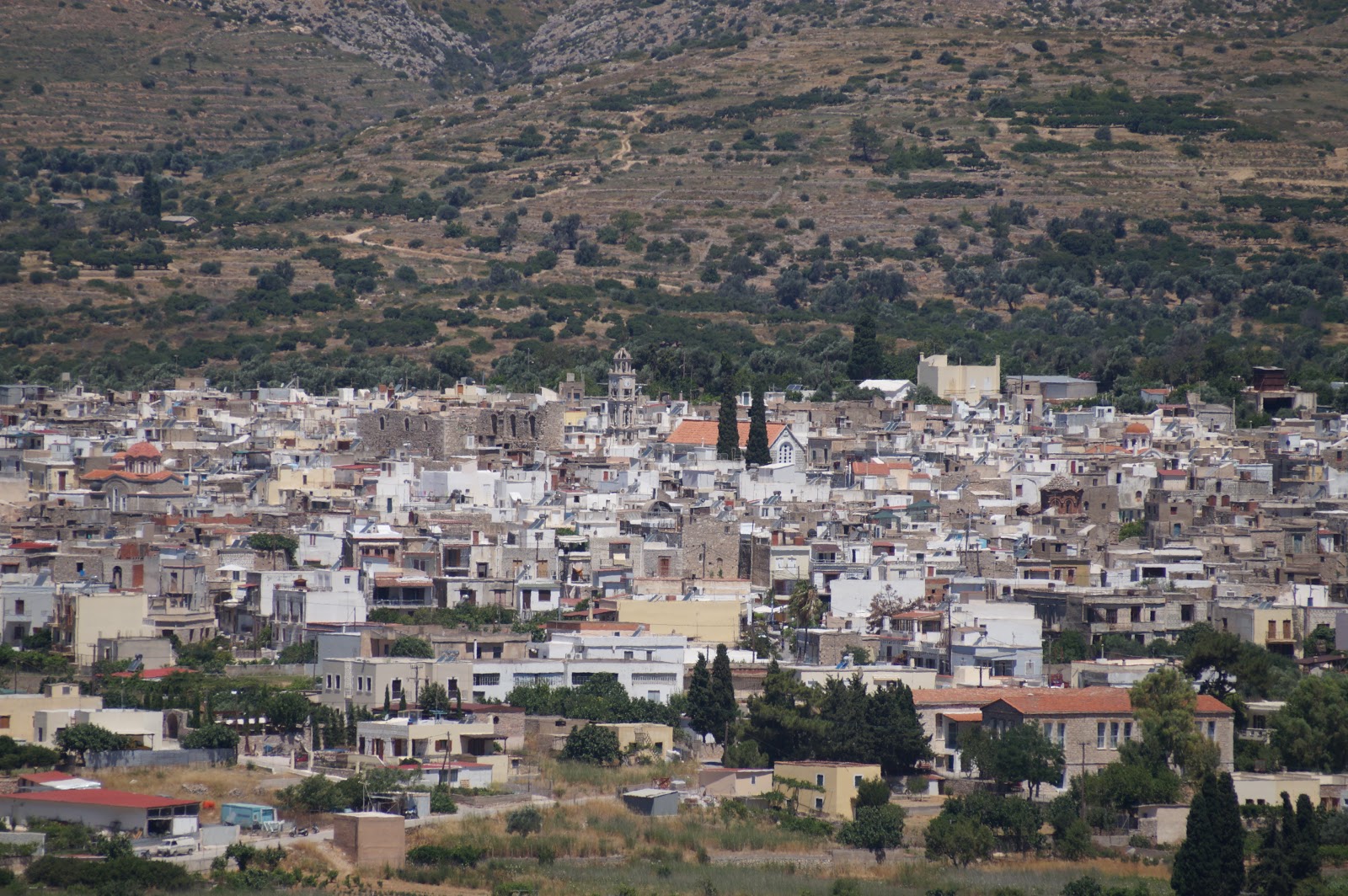
Pyrgi is the largest and most important village in the Mastichochoria. The village is known for its beauty and the intricate grey and white patterns seen on almost every building in town.  These patterns are produced by a technique called Xysta which uses cement, volcanic sand and lime to produce these wonderful patterns.

Here are some pictures from Pyrgi:


The church and the main plateiea (town square).

Here are some good examples of the xysta on the buildings around town:


 When Sue and I arrived in Pyrgi, we parked and walked to the town cemetery, seeking gravestones for George and Harikleia. We walked around the cemetery for a few minutes (I really know how to have fun on vacation), but all of the stones were much more recent.  We later learned that after 5 years, old graves are dug up and the bones moved to an ossuary (no cremation for the Orthodox). I didn't inquire at a kafeneio (my Greek is not good enough to describe what I was looking for), but we had a long discussion with the local souvenir shop owner. Neither she nor the locals in the shop knew of any Kostomenoi in town. We tried the church in town, but it was closed at mid-day so I could not ask about records.

The next day, we drove into the main town of Chios (also called Chios) and luckily we found their Town Hall (the names of the buildings were all in Greek. I had asked out hotel manager and he drew a great map of the town and showed me where the Town Hall was. He was a wonderful source of information). In the Town Hall, my Greek was good enough so that I found the records department and a young, very helpful gentleman who spoke very good English. He searched on his computer and found a Mary Kostomenos, born in 1899, but this was the person's married name. In addition, she died in 1982 (my grandmother died in 1984). It seemed that it was not the right person. I asked him to  print out the records anyway, but he said he couldn't, citing privacy policies! So, unfortunately, I could not gather any more information about the family on Chios.

Just a quick bit about masticha. Masticha was known and valued since ancient times. The sultans and rulers of the Ottoman Empire enjoyed it so much that they left southern Chios alone when they over ran and took over the island in the medieval era. The Turks left the people alone to produce masticha and took and used the yearly output of masticha for themselves. Today, masticha is used in just about everything- gum, soap, liquor, lotions, shampoo. It is also felt to have medicinal properties as well. There is a great, new Mastic Museum just outside of Pyrgi, which we visited. The views were stunning and the history of masticha was very interesting. 

Here is the museum, with Pyrgi in the background.


The lentisk tree, with surrounding white crystals. The crystals are the resin, the masticha. We would see these all around the area.


Monday, June 3, 2019

My Computer is Killing Me!


It's summer time. The weather is getting better and outdoor activities beckon. It is time to stop sitting and start working off those pounds gained over the winter. Unfortunately, sitting has reached epidemic proportions. What is contributing to the longer sitting times and what can be done to combat a sedentary lifestyle?

It seems that people are sitting more and sitting for longer periods of time. A large study compared sitting times from 2001 to 2016. During that time, the total time spent sitting in a day increased by one hour per day, in both adolescents and adults. Total sitting in time in adolescents went from 7 hours per day to 8.2 hours per day while adults went from 5.5 hours per day to 6.4 hours per day. In breaking down the cause for the increase, the researchers found that the time watching television or videos was the same, about 2 hours per day. However, the time spent sitting in front of a computer during free time, not for work or for school, rose significantly. In 2001, 43% of children and 29% of adults used a computer for more than one hour per day. By 2016 those numbers rose to 56% in children and 50% in adults.  

All of that computer clicking can be deadly. There is an increased risk for death and heart disease in those who sit for prolonged periods. This risk is even higher if those who sit many hours per day don’t do the recommended weekly activity (the recommended amount of activity is 150 minutes per week for moderate exercise, 75 minutes per week for vigorous exercise). For perspective, 65% of the US population report spending less than 150 minutes per week in leisure time activity. The combination of sitting and no activity is especially deadly. How much activity is needed to offset the risk of prolonged sitting? Meeting the 150-minute/week threshold reduces the risk and the more activity, the lower the risk for death and heart disease.  However, for those who typically sit 8 or more hours a day, at least 300 minutes per week of activity are needed to reduce the risk. Therefore, to lower the risk for heart disease and dying, less sitting and more physical activity is needed. In a week filled with work and home obligations, how can that be achieved? Here are some possible strategies.

There is a whole chunk of the day that is committed to commuting. The average commuting time in the US was 27 minutes one way in 2017 with larger cities having longer commute times (the average commute time in the New York City area is 38 minutes one way). For most people that time is spent sitting in a car. Changing commuting habits can help meet or exceed the recommended physical activity goals. It has been shown that walking or biking to work can lower the risk for heart disease by 11% and the risk for dying by 30%. If you must use a computer for work, or you like to spend your free time on computer, try a stand up desk. Better yet, put a treadmill under the stand up desk and do some walking while you do your clicking.  This would go along way towards hitting that weekly physical activity target. If you have a dog at home, take the dog out for walks! Studies have shown that dog walkers are four times more likely to reach the weekly physical activity goal than people who do not have dogs. On average, dog owners do more physical activity than non-dog owners; about 200 minutes more per week! In addition, dog owners (and especially dog walkers) have a lower risk for heart attack and heart deaths. If you can’t walk or bike to work and don’t have a dog, try doing a simple, low-cost exercise.  A study showed that young men who could complete 40 pushups had a lower risk for heart disease than those who could do 10 or less pushups.  

If you don’t have a dog and don’t like to do pushups, start a walking regimen.  A goal of 10,000 steps per day is thought to be associated with good health. Why 10,000 steps? How was that number derived? What is the data?  Despite the fact that 10,000 steps per day is touted in the media and is the goal set on wearable devices and smartphones, no one really knows where the number came from.  It is thought to have originated in Japan in the 1960’s by a company trying to promote their pedometers, but there was no hard data to support it.  More recently, the Women’s Health Study (16,000 women, average age 72 years) was able to provide some clarity.  In the study, women who took 4400 steps per day had a lower death rate than those who walked less (2700 steps per day or less), For every additional 1000 steps per day, the risk decreases by about 10%. This leveled off at about 7500 steps per day. This means that the minimum number of steps per day needed to lower the risk for death and heart disease is 4400 steps per day and there is no further benefit beyond 7500 steps per day.  How does this fit into the goal of 150 minutes per week of activity?  Calculations show that about 7000 steps per day may be sufficient to achieve the 150-minute per week goal. 

So, stop sitting. Stop clicking. Start biking to work. Or walking the dog. Or doing pushups. Or start a walking program with a goal of at least 4400 steps per day and ideally 7500 steps or more per day.  Remember, however, that if you sit for 8 hours a day, a higher number of steps, about 14,000 steps per day, are needed to offset the detrimental effects of prolonged sitting. Don’t let your computer do you in!